Human Back Bones Diagram - Human Back Bones Back Of Human Skeleton Dk Find Out - This article looks at the anatomy of the back, including bones, muscles, and nerves.
byAdmin•
0
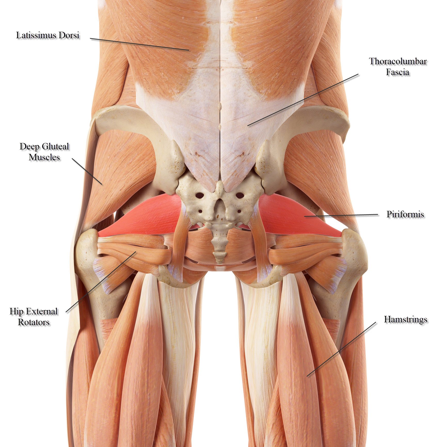
Human Back Bones Diagram - Human Back Bones Back Of Human Skeleton Dk Find Out - This article looks at the anatomy of the back, including bones, muscles, and nerves.. In addition, the broad hip bones provide protection to the delicate internal organs of the pelvis, such as the intestines, urinary bladder, and uterus. The atlas is the topmost vertebra, and along with c2, forms the joint connecting the skull and spine. Back bones diagram, human back bones skeleton, human back muscles and bones, human backbone structure, pictures of human back bones, bone. The spinal cord begins at the base of the brain and extends into the pelvis. The red lines point individual bones and the names are writen in singular, the blue lines conect to group of bones and are in plural form.
The top edge of the manubrium has a depression called the suprasternal or jugular notch. Flat bones follow the process of intramembranous ossification where the young bones grow from a primary ossification center in fibrous membranes and leave a small region of. It also covers some common conditions and injuries that can affect the back. Many of the nerves of the peripheral nervous system, or pns, branch out from the spinal cord and travel to various. The breadth of the back is created by the shoulders at the top and the pelvis at the bottom.
Spinal Anatomy Center Cervical Thoracic And Lumbar Spine Info from www.spineuniverse.com This shopping feature will continue to load items when the enter key is pressed. Pelvic bone labeled 12 photos of the pelvic bone labeled pelvic bone labeled, pelvic bone labeling quiz, pelvic bone with labeling, pelvic girdle bone labeling quiz, pubic bone labeled, bone, pelvic bone labeled, pelvic bone labeling quiz, pelvic bone with labeling, pelvic girdle bone labeling quiz, pubic bone labeled The number of bones in the human body at birth is 300. Related posts of female body back side anatomy skeleton bones diagram. This article looks at the anatomy of the back, including bones, muscles, and nerves. We are pleased to provide you with the picture named anatomy of back muscles diagram.we hope this picture anatomy of back muscles diagram can help you study and research. The lumbar spine connects to the thoracic spine above and the hips below. It is the surface of the body opposite from the chest and the abdomen.the vertebral column runs the length of the back and creates a central area of recession.
Pelvic bone labeled 12 photos of the pelvic bone labeled pelvic bone labeled, pelvic bone labeling quiz, pelvic bone with labeling, pelvic girdle bone labeling quiz, pubic bone labeled, bone, pelvic bone labeled, pelvic bone labeling quiz, pelvic bone with labeling, pelvic girdle bone labeling quiz, pubic bone labeled
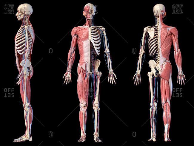
Bone names in the human body 12 photos of the bone names in the human body bone names for the human body, name all the bones in the human body quiz, name the 206 bones in the human body game, name the bones in the human body game, name three bones in the human body, … The sternum is a flat bone that is made up of three parts, the (1) manubrium, (2) body, and the (3) xiphoid process. And coccygeal the tail bone. They also provide for the attachment of muscles, and help us move around. The human skeleton, like that of other vertebrates, consists of two principal subdivisions, each with origins distinct from the others and each presenting certain individual features.these are (1) the axial, comprising the vertebral column—the spine—and much of the skull, and (2) the appendicular, to which the pelvic (hip) and pectoral (shoulder) girdles and the bones and cartilages of the. Diagram of a human female skeleton, back view. Muscle or tendon injuries can occur anywhere in the body. This article looks at the anatomy of the back, including bones, muscles, and nerves. It provides a basic framework in form of skeleton on which everything is else is laid on and anchored to. Human back bones diagram poster 28 inch x 24 inch 16 inch x 13 inch. Related posts of human back bones diagram pelvic bone labeled. Its appearance is different from the other spinal vertebrae. Related posts of human anatomy female lower back muscle anatomy triceps.
It contains the osteology, arthrology and myology of the spine and back. The human back, also called the dorsum, is the large posterior area of the human body, rising from the top of the buttocks to the back of the neck. The top edge of the manubrium has a depression called the suprasternal or jugular notch. The lumbar spine connects to the thoracic spine above and the hips below. The vertebrae, which stack like spools of thread, support the back and protect the spinal cord.
Lower Back Muscle Anatomy And Low Back Pain from ix-cdn.b2e5.com Immune and lymphatic systems of the lower torso. Our latest youtube film is ready to run. Muscle or tendon injuries can occur anywhere in the body. The breadth of the back is created by the shoulders at the top and the pelvis at the bottom. And coccygeal the tail bone. Related posts of female body back side anatomy skeleton bones diagram. The spine diagram the spine diagram shown below, consists of many bones or vertebrae,soft discs,the spinal cord, and spinal nerves. It also covers some common conditions and injuries that can affect the back.
We are pleased to provide you with the picture named anatomy of back muscles diagram.we hope this picture anatomy of back muscles diagram can help you study and research.
The spinal cord begins at the base of the brain and extends into the pelvis. These aspects are the bones of the diagram. This shopping feature will continue to load items when the enter key is pressed. Many muscles that move the trunk and legs, such as our abdominal muscles, attach to the hip bones. In the centre of your chest there is a strong bone called the sternum. This human anatomy module is composed of diagrams, illustrations and 3d views of the back, cervical, thoracic and lumbar spinal areas as well as the various vertebrae. The pelvis at the bottom of the back and the shoulders at the top of the back give the back its breadth, and it narrows in between these two regions. The human skeleton, like that of other vertebrates, consists of two principal subdivisions, each with origins distinct from the others and each presenting certain individual features.these are (1) the axial, comprising the vertebral column—the spine—and much of the skull, and (2) the appendicular, to which the pelvic (hip) and pectoral (shoulder) girdles and the bones and cartilages of the. The vertebrae, which stack like spools of thread, support the back and protect the spinal cord. Can you feel the bumps of your vertebrae along your back? As a person ages, these bones grow together and fuse into larger bones, leaving adults with only 206 bones. The atlas is the topmost vertebra, and along with c2, forms the joint connecting the skull and spine. The spine anatomy is a complex structure.
Many of the nerves of the peripheral nervous system, or pns, branch out from the spinal cord and travel to various. The neck (cervical) and low back (lumbar) regions have a slight concave curve, and the thoracic and sacral regions have a gentle convex curve (fig. It contains the osteology, arthrology and myology of the spine and back. At birth, the skeleton of a newborn has more than 300 bones; Can you feel the bumps of your vertebrae along your back?
Humans Stock Photos Offset from ak.picdn.net The curves work like a coiled spring to absorb shock, maintain balance, and allow range of motion throughout the spinal column. The first seven bones (vertebrae) of your spine form your neck. The atlas is the topmost vertebra, and along with c2, forms the joint connecting the skull and spine. Female reproductive organs of the lower torso. See lumbar spine anatomy diagram stock video clips. The bone found at the back and base of the skull. Related posts of female body back side anatomy skeleton bones diagram. These bones are connected at the back with specialized joints.
The human skeleton, like that of other vertebrates, consists of two principal subdivisions, each with origins distinct from the others and each presenting certain individual features.these are (1) the axial, comprising the vertebral column—the spine—and much of the skull, and (2) the appendicular, to which the pelvic (hip) and pectoral (shoulder) girdles and the bones and cartilages of the.
The bone found at the back and base of the skull. The atlas is a ring of bone made up of two lateral masses joined at. Diagram of a human female skeleton, back view. Many muscles that move the trunk and legs, such as our abdominal muscles, attach to the hip bones. Female reproductive organs of the lower torso. The bones provide a structural framework and protection to the soft organs. The pelvis at the bottom of the back and the shoulders at the top of the back give the back its breadth, and it narrows in between these two regions. Many of the nerves of the peripheral nervous system, or pns, branch out from the spinal cord and travel to various. The number of bones in the human body at birth is 300. Find the perfect human back. The human back, also called the dorsum, is the large posterior area of the human body, rising from the top of the buttocks to the back of the neck. The neck (cervical) and low back (lumbar) regions have a slight concave curve, and the thoracic and sacral regions have a gentle convex curve (fig. Cervical bones diagram 12 photos of the cervical bones diagram cervical bones diagram, cervix anatomy diagram related posts of human back bones diagram human bone parts name.
The muscles of the lower back help stabilize, rotate, flex, and extend the spinal column, which is a bony tower of 24 vertebrae that gives the body structure and houses the spinal cord back bones diagram. Immune and lymphatic systems of the lower torso.