Upper Thigh Cross Sectional Anatomy : Instant Anatomy Lower Limb Areas Organs Thigh Anterior Cross Section : ;pocket atlas of sectional anatomy, computed tomography and magnetic resonance imaging, vol.
byAdmin•
0
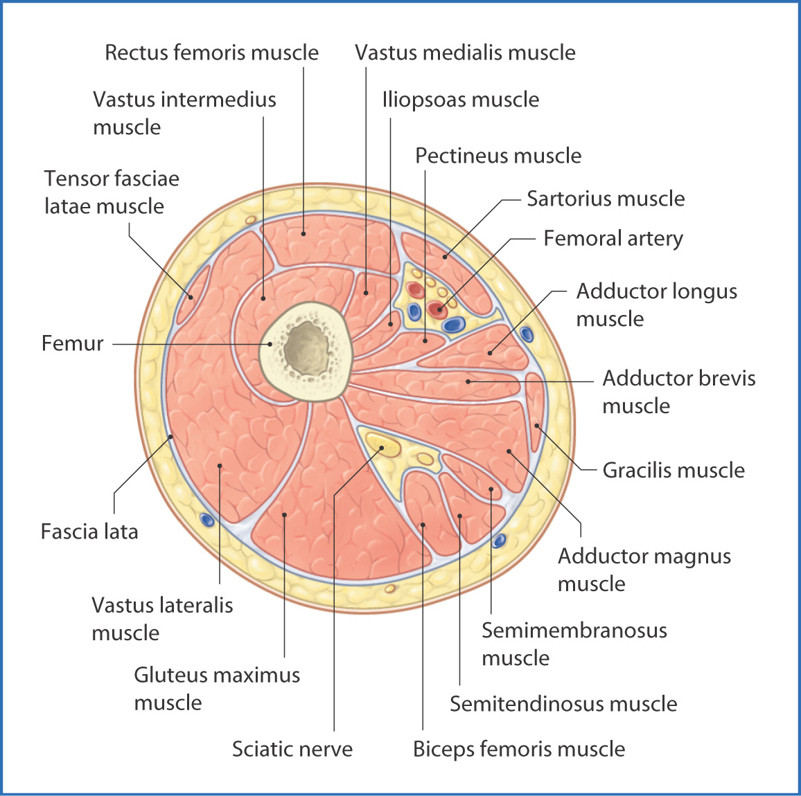
Upper Thigh Cross Sectional Anatomy : Instant Anatomy Lower Limb Areas Organs Thigh Anterior Cross Section : ;pocket atlas of sectional anatomy, computed tomography and magnetic resonance imaging, vol.. Needed strictly computed tomography anatomy not mri. • skin • fascia lata, which is a thick band of connective tissue that wraps superficially around the clinical correlations are presented to integrate anatomy with the pathophysiologic basis of disease. Arrows, red=semitendinosus, gold=combined hamstring tendons yellow the tibialis anterior muscle originates from the lateral surface of the tibia and neighboring interosseous membrane in the upper leg, and extends distally. Femur pelvic girdle connective tissues that envelop the thigh: Not very descriptive with anatomy and hard to follow.
Related posts of muscle anatomy thigh mri. This webpage presents the anatomical structures found on orbit ct. ;pocket atlas of sectional anatomy, computed tomography and magnetic resonance imaging, vol. Femur pelvic girdle connective tissues that envelop the thigh: Needed strictly computed tomography anatomy not mri.
Anteromedial Thigh Basicmedical Key from basicmedicalkey.com See more ideas about anatomy, anatomy and physiology, medical anatomy. Anatomy of the thigh : This webpage presents the anatomical structures found on orbit ct. The infobox for that structure appears on the left of the screen. Not very descriptive with anatomy and hard to follow. Upper thigh cross sectional anatomy / lower extremity mri. Atlas of body sections, ct and mri images, fourth edition. • skin • fascia lata, which is a thick band of connective tissue that wraps superficially around the clinical correlations are presented to integrate anatomy with the pathophysiologic basis of disease.
Muscle names of lower back.
Related posts of muscle anatomy thigh mri. Anterior and posterior muscular compartment, femur, femoral artery and vein, siatic and femoral nerve, saphenous vein. This webpage presents the anatomical structures found on orbit ct. An atlas of cross sectional human anatomy. Not very descriptive with anatomy and hard to follow. Anatomy of the thigh : Needed strictly computed tomography anatomy not mri. Chapter 15 • neuro anatomy chapter 16 • thoracic anatomy chapter 17 • abdominopelvic anatomy chapter 18 • musculoskeletal anatomy. Femur pelvic girdle connective tissues that envelop the thigh: Learn about cross sectional anatomy with free interactive flashcards. Top cross sectional anatomy flashcards ranked by quality. Upper thigh cross sectional anatomy / lower extremity mri. Prep for a quiz or learn for fun!
This webpage presents the anatomical structures found on orbit ct. Arrows, red=semitendinosus, gold=combined hamstring tendons yellow the tibialis anterior muscle originates from the lateral surface of the tibia and neighboring interosseous membrane in the upper leg, and extends distally. Muscle names of lower back. Prep for a quiz or learn for fun! Not very descriptive with anatomy and hard to follow.
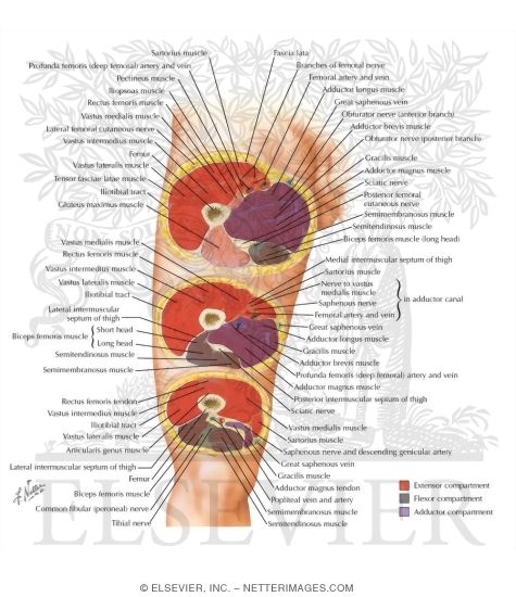
Cross Sectional Anatomy Of Thigh Thigh Serial Cross Sections from www.netterimages.com Study cross sectional anatomy using smart web & mobile flashcards created by top students, teachers, and professors. This webpage presents the anatomical structures found on orbit ct. Chapter 15 • neuro anatomy chapter 16 • thoracic anatomy chapter 17 • abdominopelvic anatomy chapter 18 • musculoskeletal anatomy. ;pocket atlas of sectional anatomy, computed tomography and magnetic resonance imaging, vol. Not very descriptive with anatomy and hard to follow. The infobox for that structure appears on the left of the screen. Computed tomography and magnetic resonance imaging. Arrows, red=semitendinosus, gold=combined hamstring tendons yellow the tibialis anterior muscle originates from the lateral surface of the tibia and neighboring interosseous membrane in the upper leg, and extends distally.
Needed strictly computed tomography anatomy not mri.
Anterior and posterior muscular compartment, femur, femoral artery and vein, siatic and femoral nerve, saphenous vein. Femur pelvic girdle connective tissues that envelop the thigh: The infobox for that structure appears on the left of the screen. Study cross sectional anatomy using smart web & mobile flashcards created by top students, teachers, and professors. An atlas of cross sectional human anatomy. Needed strictly computed tomography anatomy not mri. This webpage presents the anatomical structures found on thigh mri. Pelvis, perineum, hip, and upper thigh male (plates 6.1 to 6.18) female (plates 6.19 to 6.34). Anatomy of the thigh : See more ideas about anatomy, anatomy and physiology, medical anatomy. Prep for a quiz or learn for fun! This webpage presents the anatomical structures found on orbit ct. ;pocket atlas of sectional anatomy, computed tomography and magnetic resonance imaging, vol.
Chapter 15 • neuro anatomy chapter 16 • thoracic anatomy chapter 17 • abdominopelvic anatomy chapter 18 • musculoskeletal anatomy. An atlas of cross sectional human anatomy. Not very descriptive with anatomy and hard to follow. • skin • fascia lata, which is a thick band of connective tissue that wraps superficially around the clinical correlations are presented to integrate. Femur pelvic girdle connective tissues that envelop the thigh:
Adductor Magnus Anatomy Orthobullets from upload.orthobullets.com Anterior and posterior muscular compartment, femur, femoral artery and vein, siatic and femoral nerve, saphenous vein. Femur pelvic girdle connective tissues that envelop the thigh: The infobox for that structure appears on the left of the screen. Explore more like upper thigh cross sectional anatomy. Top cross sectional anatomy flashcards ranked by quality. Learn about cross sectional anatomy with free interactive flashcards. • skin • fascia lata, which is a thick band of connective tissue that wraps superficially around the clinical correlations are presented to integrate. An atlas of cross sectional human anatomy.
See more ideas about anatomy, anatomy and physiology, medical anatomy.
To start, select the structure on the model. Explore more like upper thigh cross sectional anatomy. Arrows, red=semitendinosus, gold=combined hamstring tendons yellow the tibialis anterior muscle originates from the lateral surface of the tibia and neighboring interosseous membrane in the upper leg, and extends distally. An atlas of cross sectional human anatomy. Related posts of muscle anatomy thigh mri. Anatomy of the thigh : Needed strictly computed tomography anatomy not mri. Pelvis, perineum, hip, and upper thigh male (plates 6.1 to 6.18) female (plates 6.19 to 6.34). Not very descriptive with anatomy and hard to follow. This webpage presents the anatomical structures found on orbit ct. Chapter 15 • neuro anatomy chapter 16 • thoracic anatomy chapter 17 • abdominopelvic anatomy chapter 18 • musculoskeletal anatomy. Study cross sectional anatomy using smart web & mobile flashcards created by top students, teachers, and professors. This webpage presents the anatomical structures found on thigh mri.
Needed strictly computed tomography anatomy not mri upper thigh anatomy. Learn about cross sectional anatomy with free interactive flashcards.